http://www.youtube.com/watch?v=bCPxwSSv5i0
http://www.youtube.com/watch?v=b34PHY2RPGQ
http://www.youtube.com/watch?v=CZPeBhVNAtY
Well her we go again off to the vet to get Gracys evaluation and first treatment for her heart worms. It was a long day of waiting and praying for the best outcome. At 4;00 pm we received a call from the doc, we can go there to pick her up. I know Terri was relieved because we thought she needed to stay overnight, doc said" not yet, latter on she will need to be watched overnight".
She started her first treatment which was a series of blood test to check all of her organs to see if they are healthy enough to continue and a Xray view of her lungs and heart for diagnoses of heart worm phase, which will determine what path to take in treatment. Then doc started her on Triheart Plus 1-25# to kill any larva and to give treatment better chance for success, Gracey will be on heart pill for 2 months before 1st Immiticide treatment in January.
Here is what the Doc said " Bright, alert and responsive; good body condition, Cardiovascular Regular rhythm; no murmur detected; strong femoral pulses;CRT<>
Heart worm disease classification 2.
http://www.youtube.com/watch?v=P6F9KApqkII
http://www.youtube.com/watch?v=djzpi4msUd8
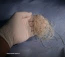
We were relieved to here it was only in phase 2, she still has an uphill battle but there is a 98% success rate. We looked up some info at http://www.heartwormsociety.org/ and here is some of the facts we found.
(X-ray)
Radiographic abnormalities develop early in the course of the disease. X-rays of the heart and lungs are the best tools available to evaluate the severity of the disease and to develop a prognosis. Typical changes observed are enlargement of the following structures: pulmonary arteries in the lobes (particularly the lower lobes) of the lung, main pulmonary artery, and right side of the heart. Blunting and thickening, usually along with tortuosity (abnormal twists or turns), of pulmonary arteries, is often noted. Inflammation is often found in the lung tissue, particularly the tissue that surrounds the pulmonary arteries.

http://www.youtube.com/watch?v=sz3xs7czgcw
Examination
A heartworm infected dog with mild disease may appear to be perfectly normal upon physical examination. Severely affected dogs, however, may show signs of right-sided heart failure. Labored breathing or crackles may be heard in the lungs due to vascular clots and elevated pressure. A history of coughing and inability to exercise are among the earliest detectable abnormalities. Tachycardia (rapid heartbeat), ascites (fluid in the abdomen) and hepatomegaly (enlarged liver) indicate right-sided congestive heart failure. Hemoptysis (blood in the sputum) occasionally occurs and indicates severe clots and complications within the lungs. Anorexia (loss of appetite), cachexia (severe weight loss), syncope (fainting) jaundice or yellow bile pigmentation present in the skin and mucus membranes may appear in severely affected dogs. Occasionally, heartworms are reported in abnormal locations such as the eyes, abdominal cavity, cerebral artery and spinal cord. Clinical signs and disease experienced in such infections depend largely on the location of the worms. The primary response to the presence of heartworms in dogs, however, occurs in the heart and lungs.
http://www.youtube.com/watch?v=VOLzFsNOJ-4


No comments:
Post a Comment